Table Of Content

LOS ANGELES, CALIFORNIA — The capacity for hearing recovery remains elusive in adults with hearing loss due to the non-regenerative nature of the sensory hearing cells within the inner ear. In another study from March 2013, a research team honed in on the gene Tsc2 in Purkinje cells of the cerebellum and found that a loss of Tsc2 in Purkinje cells lead to autistic-like behavioral deficits. In 2007, my father died unexpectedly of a heart attack leaving his quest to find some type of "holy grail" in neuroscience incomplete. I made a vow at his funeral that I would pick up the torch and try to find answers to his hypotheses about Purkinje cells and the cerebellum. Every morning, I wake up hoping there will be new research that helps to decrypt the mysteries of the cerebellum. Needless to say, I was thrilled to read about the new study on eye movements and Purkinje cells released this morning.
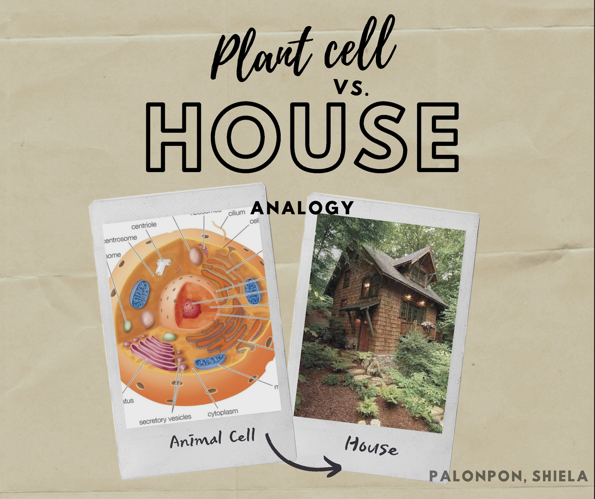
Do All Cells Contains the Same Number of Mitochondria?
(a) The mitochondrial outer membrane is the site of important signalling events during the innate immune response. Detection of viral nucleic acids by Rig-like receptors (RLRs) induces dimerization of MAVS, a protein of the mitochondrial outer membrane. Dimerized MAVS recruits signalling adaptors that initiate downstream activation of IRF3/7 and NF-κB, transcription factors that induce expression of type I interferons and pro-inflammatory cytokines. MAVS is regulated by NLRX1, a protein which downregulates MAVS when localized to the outer membrane, but activates MAVS when at the inner membrane by interacting with Complex III to induce ROS production. Release of mtDNA during infection can also activate the NLRP3 inflammasome.
AnalysisSportThe science behind Zone 2 training for athletes
The addition of the acetyl group to oxaloacetate forms citrate and the cycle repeats. The breakdown of citrate into oxaloacetate releases a further two CO2 molecules and one molecule of ATP (through substrate-level phosphorylation). The number of mitochondria per cell varies widely—for example, in humans, erythrocytes (red blood cells) do not contain any mitochondria, whereas liver cells and muscle cells may contain hundreds or even thousands. The only eukaryotic organism known to lack mitochondria is the oxymonad Monocercomonoides species.
The powerhouse function
They found that the size ratio between the algae and UCYN-A bacterium remains similar across different species related to the B. The growth appears to be controlled by an exchange of key nutrients, linking up their metabolisms. This synchronization of growth rates led the researchers to call UCYN-A organelle-like. Nitrogen is a very important nutrient for life to exist and plants normally get it through mutual relationships with the bacteria that remain separate from the plant or algae. Bigelowii algae had this kind of symbiotic relationship with a bacterium called UCYN-A. Endosymbiosis where the host life form becomes fundamental to another organism’s function has only happened three known times.
Study Casts Doubt on One of Our Key Assumptions About The 'Powerhouse of The Cell' - ScienceAlert
Study Casts Doubt on One of Our Key Assumptions About The 'Powerhouse of The Cell'.
Posted: Wed, 16 Oct 2019 07:00:00 GMT [source]
The citric acid cycle results in the formation of NADH (from NAD+) which transports electrons to the final stage of cellular respiration. ATP is produced via chemiosmosis, a process that also occurs in the inner membrane of the mitochondrion. Chemiosmosis involves the transmembrane protein ATP synthase which produces ATP from ADP and inorganic phosphate. ATP synthase uses the concentration gradient of hydrogen ions to drive the formation of ATP. As the electrons move through the electron transport chain, hydrogen ions are pushed out into the intermembrane space, producing a higher concentration of H+ outside the membrane. The consumption of H+ through incorporation into water molecules further increases the concentration gradient.
Outer membrane
As mitochondrial protein import and oxidative metabolism can be hijacked by virulence factors [112], these interactions may make MAVS sensitive to consequences of infection. Finally, if mitochondria are compromised by infection, the increased ROS and release of mtDNA into the cytosol can activate the NLRP3 inflammasome to evoke an inflammatory response [113,114] (figure 4a). Most of the proteins and other molecules that make up mitochondria originate in the cell nucleus.
During mitosis, a highly fused and reticular mitochondrial network progressively fragments to small tubular organelles that segregate in anticipation of cytokinesis [127,131]. Mitochondria can also delay cell-cycle progression to increase their biogenesis [132], because of insufficient nucleotide production [133], or because of ROS accumulation [134]. Moreover, the fusion mediator Mfn2 can sequester both Ras and Raf to inhibit proliferative signalling [135]. The outer mitochondrial membrane is permeable to Ca2+, in part due to channel-forming VDAC proteins [87] and export via SLC8A3 [88]. The mitochondrial inner membrane calcium uniporter (MCU) complex regulates transport into the matrix (figure 3c). Permeability of the MCU complex is calibrated by two regulatory subunits, MICU1 and MICU2, that are linked by an intermolecular disulfide bond introduced by hMia40 [89,90].
Although free radicals are damaging, they have an important signalling role. They are unique organelles present in almost all eukaryotic cells that are responsible for generating the cell’s supply of adenosine triphosphate (ATP), the energy currency of the cell. And, although they are popularly referred to as the powerhouse of the cell, they carry out a wide range of actions that are much less known about.
DNA repair
Mitochondria were essential for the development of life as we know it due to their multiple functions, especially ATP production. New stress responses are emerging that demonstrate the reciprocal communication between mitochondria and cytoplasm. Cerevisiae activates the proteasome to mitigate mitochondrial precursor accumulation in the cytosol [187]. Cerevisiae, the proteasome is also engaged by Ubx2 to clear mitochondrial protein precursors arrested during translocation, blocking the TOM complex [191]. Reciprocally, mitochondria can degrade defective proteins to aid cytosolic proteostasis. Intriguing for further research are reports of lysosomal fusion of mitochondria-derived vesicles enriched for non-natively oxidized proteins [194,195] and the extracellular jettison of aggregates by neurons of C.
(b) Mitophagy is a process that allows damaged mitochondria to be identified and destroyed. Under normal conditions, PINK1 is imported into mitochondria and degraded by PARL. When mitochondria are damaged, import is impaired and PINK1 accumulates in the TOM complex at the outer membrane. Autophosphorylated and active PINK1 at the outer membrane phosphorylates monoubiquitin molecules on outer membrane proteins, recruiting and activating the E3 ubiquitin ligase Parkin. Activated Parkin synthesizes polyubiquitin chains that recruit autophagy receptors to initiate mitophagy.
Acetyl-CoA is the primary input for the TCA cycle, and can be obtained through metabolism of glucose, fatty acids and amino acids. Electrons are subsequently transferred from NADH and FADH2 onto Complexes I and II of the electron transport chain. Electrons are passed through Complexes III and IV, which transport protons into the intermembrane space. Protons are allowed to flow back into the matrix through ATP synthase (Complex V), which uses the energy of the proton gradient to convert ADP to ATP.
Additionally, mitochondrial Ca2+ regulation influences hormone secretion [100], tissue regeneration [101] and interferon-β signalling via the mitochondrial antiviral signalling protein, MAVS [102]. The outer mitochondrial membrane is freely permeable to small molecules and contains special channels capable of transporting large molecules. In contrast, the inner membrane is far less permeable, allowing only very small molecules to cross into the gel-like matrix that makes up the organelle’s central mass. Stem cell differentiation also relies on mitochondria as a ‘metabolic switch’.

No comments:
Post a Comment